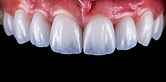
Dental erosion is a common form of non-carious tooth wear and may progress for a long time before a patient becomes aware of it.
Unlike abrasion or attrition, erosion is driven primarily by chemical exposure to acids rather than direct mechanical wear. Acid softens the tooth surface, after which normal brushing, chewing or grinding can contribute to further loss. Early erosion can be symptom-free.
Dental erosion describes loss of enamel and dentine through non-bacterial acid exposure. Acids can come from outside the body (extrinsic) or from gastric contents (intrinsic), and mechanical wear often acts together with the chemical process.
Common extrinsic sources include:
frequent consumption of soft drinks, energy drinks, fruit juices, sports drinks, citrus products, wine and other acidic foods or beverages. Repeated small sips throughout the day and reduced salivary flow increase the duration of acid contact.
Intrinsic acid exposure may occur when gastric acid reaches the mouth, for example with gastro-oesophageal reflux, recurrent vomiting or eating disorders. The pattern of wear can give clues to the source, but medical assessment is required when an underlying condition is suspected.
Erosive tooth wear can produce smooth, rounded or cupped surfaces, loss of natural tooth contours and increasing sensitivity. Incisal edges may appear thinner or more translucent. With advanced generalised wear, tooth height and restorative space may also be affected.


If substantial tooth height is lost, the lower facial third can appear shorter and lip or cheek support may change. Facial folds and wrinkles are multifactorial, however, and should not be attributed to dental erosion alone.

Loss of vertical tooth height
Without significant loss of vertical dimension:

With reduced vertical dimension:

Edentulous patient:

Diagnosis
At our practice in Basel, assessment includes clinical examination, photographic documentation, digital intraoral scans and a detailed dietary and medical history. If reflux or recurrent vomiting is suspected, medical assessment may be recommended. Diagnosis is based primarily on the characteristic pattern and progression of tooth-surface changes.
Treatment of erosive tooth wear has two main goals:
reduce further acid exposure and restore lost tooth structure only when clinically necessary.
Important preventive measures can include:
identifying and reducing repeated exposure to acidic drinks or foods
changing drinking habits, for example avoiding prolonged sipping and frequent acid contact
rinsing with water after acid exposure and allowing saliva time to neutralise the oral environment
using fluoride and other evidence-based preventive measures according to individual risk
adjusting dietary habits without unnecessarily restricting a balanced diet
medical assessment when reflux or recurrent vomiting is suspected
After significant acid exposure, it is generally sensible to avoid immediate aggressive toothbrushing and allow time for saliva to neutralise and reharden the surface. Practical advice should be adapted to the person's overall caries and erosion risk rather than relying on one rigid waiting time.
Restorative options depend on severity:
additive composite restorations for selected early or moderate structural loss
ceramic veneers, partial-coverage restorations or crowns when more extensive reconstruction is required
reconstruction of the vertical dimension in selected patients with advanced generalised tooth wear
Because erosive wear is often generalised, treatment planning should identify the cause before extensive restoration. Digital planning can help reconstruct tooth form conservatively and test changes before definitive treatment.
Frequently asked questions about dental erosion
What exactly causes dental erosion?
Repeated exposure to acids from diet or gastric contents softens the mineralised tooth surface and can lead to progressive loss of enamel and dentine.
Can erosion occur without pain?
Yes. Many patients notice sensitivity only after substantial surface changes have already occurred.
Which drinks are particularly erosive?
Many soft drinks, energy drinks, fruit juices, smoothies, sports drinks and wines are acidic. Frequency and contact time are often as important as the beverage itself.
What does “sipping” mean and why does it matter?
Repeatedly taking small sips of an acidic drink over a long period extends the time during which the teeth are exposed to a low pH.
Can chewing gum help?
Sugar-free chewing gum can stimulate saliva in suitable patients and may help neutralise acids. It is an adjunct, not a substitute for reducing repeated acid exposure.
Can eroded enamel grow back?
A softened surface can reharden to some extent through remineralisation, but tooth structure that has been physically lost does not regenerate. Restorative replacement is considered only when function, sensitivity, structure or aesthetics justify it.
What should I do if reflux is causing erosion?
Medical assessment is important because controlling the underlying reflux or vomiting is central to preventing continued acid exposure. Dental protection alone does not address the cause.
If your teeth look thinner, smoother or more translucent, or sensitivity is increasing, a structured assessment can determine whether erosion is active, what is driving it and whether preventive monitoring or restorative treatment is appropriate.
References
Lussi A, Jaeggi T. Erosion—diagnosis and risk factors. Clin Oral Investig. 2006;10 Suppl 1:S35–S39. PMID: 16687883.
Lussi A, Schlueter N, Rakhmatullina E, Ganss C. Dental erosion – an overview with emphasis on chemical and histopathological aspects. Clin Oral Investig. 2011;15(6):755–760. PMID: 21298338.
Mulic A, Tveit AB. Risk indicators for dental erosive wear. Acta Odontol Scand. 2021;79(6):416–422. PMID: 33410255.
Järvinen V, Rytömaa I, Heinonen OP. Risk factors in dental erosion. J Dent Res. 1991;70(10):1534–1539. PMID: 1925485.
Mehta SB, Banerji S, Millar BJ, Suarez-Feito JM. Current concepts on the management of tooth wear: part 2—restorative management. Br Dent J. 2012;212(2):73–82. PMID: 22273708.