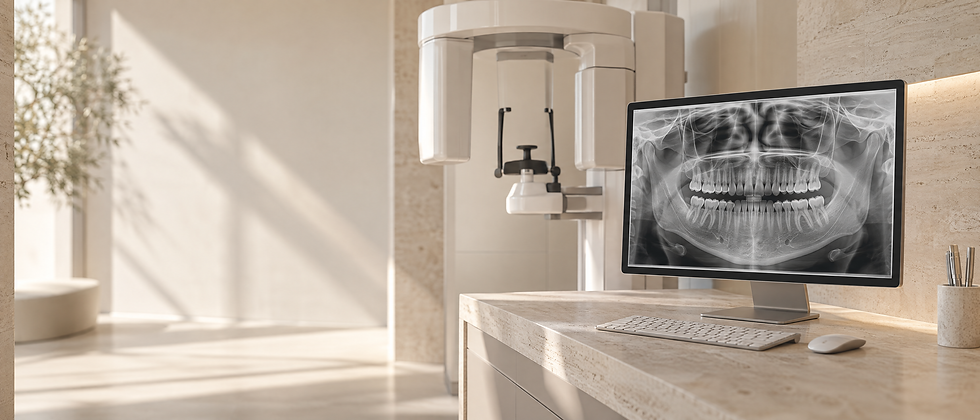
Panoramic Dental X-Ray

Panoramic Dental X-Ray (OPG) in Basel
In brief: panoramic dental X-ray (OPT/OPG)
An OPT, also called an orthopantomogram or panoramic radiograph, is a two-dimensional X-ray that shows the upper and lower jaws in a single image.ItIt is an important overview image in dentistry because it can show the teeth, jaw bones, wisdom teeth, maxillary sinuses and temporomandibular-joint regions at the same time.
In our Basel practice, we use modern digital X-ray equipment and select the imaging method according to the clinical question, following the principle of keeping radiation exposure as low as reasonably achievable.
We are members of the Swiss Society of Dentomaxillofacial Radiology (SGDMFR) and regularly update our training to maintain high standards of safety in dental radiology. https://www.sgdmfr.ch/
What can a panoramic dental X-ray show?
A panoramic X-ray provides a rapid overview of:
The condition of all teeth and tooth roots
Dental disease in areas that are difficult to assess clinically
The position of wisdom teeth and their relationship to nearby nerves
Bone loss associated with periodontitis
Cysts, inflammation or other suspicious lesions
The maxillary sinuses
The contours of the temporomandibular joints
Basic imaging for implant planning
Potential Benefits and Treatment Goals
Quick and straightforward: the scan itself usually takes only a few seconds.
Niedrige Strahlendosis dank moderner Digitaltechnik
Broad overview of both jaws
Useful diagnostic information for many treatment decisions
Particularly useful for assessing wisdom teeth, general bone levels and other broad dental findings
Valuable for implant and surgical planning
When is a panoramic X-ray useful?
Initial examination / baseline diagnostics
Assessment of wisdom teeth
Periodontal assessment
Investigation of cysts and inflammatory lesions
Screening of the temporomandibular joints
Planning before implant treatment
Monitoring during orthodontic treatment
For more demanding questions – for example implant planning, complex root anatomy or sinus involvement – CBCT may be used additionally when three-dimensional information can change the clinical decision.
Radiation protection and safety
: as low as diagnostically acceptable.
For CBCT, the smallest field of view that adequately answers the clinical question is preferred.
During pregnancy, dental radiographs are taken only when clinically justified; necessary imaging should not be withheld when the benefit outweighs the risk.
Modern digital technology helps keep radiation exposure as low as reasonably achievable.
Treatment Process
Radiation protection and positioning are adapted to current recommendations and the equipment used; routine lead aprons are no longer required for many modern dental radiographic examinations when appropriate collimation and technique are used.
the X-ray unit rotates around the head
the image is available digitally immediately
we discuss the relevant findings with you directly
Scientific Context
Panoramic radiography is an established first-line imaging method
It can provide useful information on wisdom-tooth position, periodontal bone changes and other jaw findings.
Sedaghatfar M. et al. (2019). Dentomaxillofac Radiol. PMID: 30661391
Modern digital systems can reduce radiation exposure
Digital panoramic systems generally allow lower radiation doses than older analogue techniques.
Harms H. et al. (2020). Radiat Prot Dosimetry. PMID: 32125052
Useful in periodontal assessment
Panoramic radiographs can demonstrate horizontal bone loss and contribute to periodontal diagnosis and treatment planning.
Mol A. (2004). Periodontol 2000. PMID: 15212352
Relevant for wisdom-tooth assessment
Panoramic imaging can help assess the relationship between lower wisdom teeth and the inferior alveolar nerve canal.
Rood JP. (1990). Br J Oral Maxillofac Surg. PMID: 2390438
Costs and Individual Treatment Planning
(depending on clinical indication, complexity and insurance status)
Treatment | Indicative Range |
OPT / OPG (digital panoramic X-ray) | 180 |
Assessment and discussion of findings | included / 40–80 |
Frequently asked questions
Is a panoramic X-ray harmful?
The radiation dose of a panoramic radiograph is relatively low, but it is still ionising radiation. We therefore take an image only when it is expected to provide clinically relevant information.
Does every implant case require a panoramic X-ray?
A panoramic image is often useful as baseline imaging. For precise three-dimensional implant planning, a
Can a panoramic X-ray detect tooth decay?
It may reveal some lesions, but bitewing or intraoral radiographs are generally more accurate for detecting many forms of caries.
How often can a panoramic X-ray be taken?
Only when clinically indicated; the interval depends on your findings, risk profile and treatment needs rather than a fixed schedule.
A clear overview supports safe planning
A panoramic radiograph is a quick overview examination that can support diagnosis and treatment planning. It does not replace the clinical examination and may need to be supplemented by targeted 2D or 3D imaging depending on the question.
If you would like to know whether a new panoramic X-ray is appropriate in your case, we can advise you in Basel based on your clinical findings and select the imaging method that provides the necessary information with the lowest appropriate radiation exposure.
Diagnostics and Planning
Diagnostics should answer a specific clinical question. Clinical findings, photographs, scans and radiographs are therefore combined according to the indication rather than used routinely. The result supports an understandable diagnosis, comparison of alternatives and transparent treatment planning.
Limitations, Risks and Alternatives
Every diagnostic method has limitations. Imaging does not replace the clinical examination, and additional images are useful only when they can improve a specific clinical decision. Findings are therefore interpreted together with symptoms, the clinical examination and previous imaging.
Panoramic dental X-ray in Basel: experience and clinical context
At Fachzahnarztpraxis Dr. Gasser in Basel, diagnostics are used to support structured clinical decision-making. Dr Thomas Gasser is a specialist in reconstructive dentistry (SSRD), holds an MAS in Oral Implantology (UZH) and is a member of the SGDMFR. Digital methods are used where they meaningfully complement clinical assessment and planning.More about Dr Thomas Gasser’s training and qualifications.
Related Topics on Our Website
Depending on the individual situation, CBCT,Examination or Intraoralscanner may also be relevant.You can find further dental services in Basel in our treatment overview.
Literature and Scientific Context
PubMed PMID 39225920 – Diagnostic accuracy of periapical radiography and panoramic radiography in the detection of apical periodontitis: a systematic review and meta-analysis
PubMed PMID 38534297 – The Accuracy of Bone Assessment Distal to Lower Second Molars Using Panoramic Radiography: A Systematic Review and Meta-Analysis
The selected literature provides scientific context. Which findings are relevant to a specific treatment depends on the diagnosis, the starting situation, individual risks and the treatment goals agreed together.



Other treatments